Infantile Spasms (West Syndrome)
WEST SYNDROME (INFANTILE SPASMS)
Introduction
West Syndrome is a severe epilepsy syndrome of infancy characterized by the triad of symptoms: Infantile spasms, Hypsarrhythmia EEG Pattern, Severe intellectual disability.
History
Infantile Spasms were first described by Dr. W. J West in the early 1840’s. He witnessed what he described as “Bobbing” events noting quick Tonic-Clonic movements of the head occurring in clusters over the span of three minutes.
It wasn’t until nearly 100 years later that the interictal EEG pattern termed Hypsarrhythmia would be connected with the clinical symptoms that Dr. West had described as “Infantile spasms.” The infantile spasms in conjunction with the Hypasarrhythmia EEG pattern would later be termed “West Syndrome.”
Symptoms
As mentioned previously, West Syndrome is classically characterized by the “Triad” of symptoms: Infantile spasms, Hypsarrhythmia EEG pattern, Severe intellectual disability.
Infantile Spasms
Infantile spasms are the characteristic clinical symptom observed during the Ictal period. Infantile spasms are typically broken down into two categories known as the flexor spasm and extensor spasm. The flexor spasm involves the contraction of the flexor muscles in the neck, trunk and limbs that has been described as a “Self-Hugging” motion. The extensor spasm is described as the lateral extension of the neck and truck away from the body. Infantile spasms are also known as Jackknife seizures.
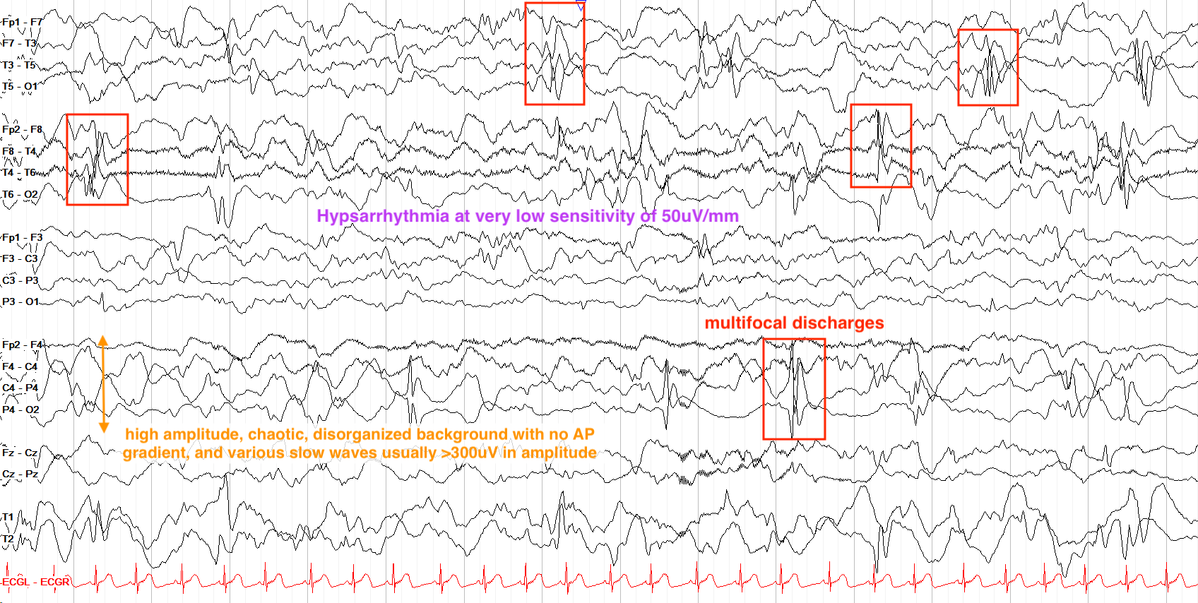
Hypsarrhythmia
The Hypsarrhythmia pattern is observed during interictal-EEG.
The Hypsarrhythmia pattern is described as a
- High voltage (>300μV)
- Multi-focal
- Slow waves with superimposed spike & wave discharges
<a title="Ralphelg, CC BY-SA 3.0 , via Wikimedia Commons” href=”https://commons.wikimedia.org/wiki/File:Hypsarrhythmia.png”>
Ralphelg, CC BY-SA 3.0, via Wikimedia Commons
Figure 1.1 Awake interictal EEG showing disorganized high amplitude spike and wave compatible with Hypsarrhythmia.
From “The Pediatric EEG”, by David Valentine M.D., 2020, (https://www.learningeeg.com/pediatric). Copyright 2020 by David Valentine
Figure 1.2 Hypsarrhythmia pattern showing multifocal discharges.
Severe Intellectual Disability
In West Syndrome, studies have shown that ~70% of patients have significant intellectual delay. Evidence suggests that longer duration of spasms (Increased seizure frequency) is correlated with worse intellectual outcomes. Even in the case of spontaneous remission of West Syndrome, developmental prognosis is often unfavorable.
Classification of disease
West Syndrome tends to have two classifications of diagnoses pertaining to the origin of disease.
Idiopathic: Suspected to account for ~9-14% of diagnosed West Syndrome. This diagnosis is only given if there is normal development prior to onset of seizures, and there is no suspected underlying disorder.
Cryptogenic: Accounts for the majority of West Syndrome diagnoses. Typically in the case of West Syndrome, this diagnosis indicates that an underlying symptomatic cause is at play, but lacks official diagnosis.
Genetic Factors: Certain genes have been identified as being contributory to West Syndrome. These include the:
- TSC 1 & 2 genes (Known to cause Tuberous Sclerosis which is an autosomal dominants disease associated with another epilepsy syndrome, Sturge-Weber)
- ARX & CDKL5 genes (Both located on the X chromosome with evidence to suggest that a higher incidence of West Syndrome is associated with these genes)
Treatments & Prognosis
Prognosis of West Syndrome is poor and directly related to the severity of disease. Severe intellectual disability is present in nearly 70% of patients. Infantile spasms rarely last until adulthood as in ~50-70% of patients develop another seizure disorder. The evolution from West Syndrome to Lennox-Gastaut syndrome being the most common evolution.
Typical anti-epileptic drugs are often ineffective in patients with West Syndrome. The complete reduction of seizures in patients with West Syndrome is rare and treatment usually aims to decrease seizure frequency but rarely aims to keep patients seizure free.
Typical medications used in the treatment of West Syndrome include ACTH, Vigabatrin, and Corticosteroids. Often times multiple medications will be used together to decrease seizure frequency.
Another common treatment in West Syndrome is the Ketogenic diet. Studies have shown that in conjunction with ACTH, the Ketogenic diet reduced seizure frequency in nearly 65% of patients.
Key Takeaways
- West Syndrome is characterized by a characteristic Hypsarrhytmia EEG pattern, Infantile spasms, and severe intellectual disability.
- The Hypsarrhytmia pattern is a high voltage, slow wave pattern with multifocal spike activity.
- The progression of West Syndrome to Lennox-Gastaut is very common.
- The Ketogenic Diet along with ACTH are the mainline treatments for West Syndrome.
Objective 2: Explain the advances made in the understanding of neuroscience in the first 5000 years of recorded history.
History of Neuroscience Objective 2 Video Lecture
The reason we don't know much about what Neolithic and Indigenous peoples knew of trepanning is that we don't have any written record of their activities: the skulls are the only evidence they left. Some of the oldest European skulls date from 10,000 years ago (8000 BCE). Writing, on the other hand, started about 3400 BCE. The word "brain" started to appear in Egyptian hieroglyphics about 3000 BCE.

One key piece of evidence for an early system of neuroscience is the Edwin Smith surgical papyrus, dating from about 1700 BCE. In this record, an unnamed Egyptian physician is studying battle wounds to the head. He notes
- pulsations of the exposed brain
- the appearance of gyri (bumps) and sulci (grooves) on the brain
- "he speaks not to thee"
- "he shudders exceedingly" (probably a description of seizures from brain injury)
This knowledge of brain function was transferred from Egypt to Ancient Greece as knowledge and civilization shifted in the ancient Western world. Alcmeon of Croton[1], writing about 480 BCE, knew that the brain contained the "governing faculty". Herophilus carried out dissections of the cerebrum and cerebellum about 300 BCE. The more famous Greek physician Galen followed his work some 500 years later. Erasistratus, working about the same time as Herophilus, performed experiments on the living brain.

Unfortunately, as we will see occurring over and over, the Greeks' understanding of neuroscience was limited by their models of natural philosophy. The Greeks believed that all bodily functions were controlled by bodily humors, a collection of four different kinds of liquid which were found in the human body. The Greeks called these choles. Not only are Shakespeare's plays from c. 1600 CE imbued with this philosophy, but the English language still carries these ideas:
- melancholy describes an excess of black bile;
- cholera is a disease where patients make a lot of greenish liquid;
- a cholecystectomy is an operation where the gall bladder (–cyst– = fluid-filled sac; –ectomy = removal) is surgically separated from the body.
Hippocrates (460–379 BCE) believed that the seat of knowledge was the brain, while Aristotle (384–322 BCE), looking at the same evidence and writing around the same time, concluded the seat of knowledge is the heart, while the brain serves to cool the blood, a sort of radiator keeping the choles from getting too hot.

While Rome became the pre-eminent seat of learning and political power in the Common Era, the physician Galen (130–200 CE), born in modern Turkey, of Greek heritage, and a Roman citizen, carried on the Greek philosophical tradition. Like Herophilus, he dissected the brain. This led to his conclusions which illustrate another recurring theme in the history of neuroscience: getting the right answer for the wrong reasons. He concluded that the cerebrum is doughy, so it must store memories, while the cerebellum is hard, like a contracted muscle, so it must control the muscles. He was unable to get past the theory of bodily humors, so he believed the ventricles (fluid-filled spaces of the brain) were reservoirs for bodily humors.
The fall of Rome, the loss of the library at Alexandria, and the subsequent so-called Dark Ages froze Western civilization's understanding of neuroscience in this state for 1500 years. But progress was still made in other places, and in some ways was more durable because it wasn't limited by the Greek insistence on the theory of bodily humors. For example, Persian Hali Abbas (علی بن عباس مجوسی) translated Galen into Arabic for his illustrated medical textbook Kitab al-Maliki (Complete Book of the Medical Art). From this scholarly work, for example, we derived the modern terms dura mater, arachnoid mater, and pia mater for the three layers of membranes covering the brain. (These are Latin words, translated poorly from the Arabic, as we will discuss elsewhere.)
Exploration and the invention of the printing press led to a political, religious, and social realignment known to the modern world as the Western Renaissance (a French word that means, literally, "re-birth"). Starting about 1420 CE, Portugese explorers begin to sail the Atlantic Ocean, with the prevailing winds and currents leading them along the African coast and around the Cape of Good Hope to the south. As they began to venture further west, they ended up conquering indigenous tribes in what is now Brazil. Similarly, Spanish explorers ended up further north. (These discoveries are reflected in the languages spoken in different parts of the so-called New World.) About the same time, in Germany, Johannes Gutenburg invented the printing press (c 1440 CE), which freed written records from having to be copied in monasteries and made learning to read accessible to the common man.
 Anatomical knowledge could now be transmitted more easily from teacher to student and from culture to culture, and Western human understanding of anatomy blossomed as a result. For example, the great Flemish anatomist Vesalius (c 1540 CE), through his incredible artistic skill, both increased our anatomical vocabulary and our anatomical knowledge.
 The highly-touted Scientific Method, which is taught to every child in school, was still in its rudimentary stages and was still (as now) limited by technology and pre-conceptions. For example, René Descartes (1596-1650 CE), perhaps the most brilliant natural philosopher of the 17th century, was still forced by the theology of the time to propose a system based on Greek humors that maintained a separation between the mind (or soul) and the body (or brain substance). The French elites of Descartes' time were enamored of statuary that operated on the principle of hydraulics (moving water under different pressures). So of course it was obvious that when the foot is exposed to a fire, there must be movement of fluid in a series of channels which then end up causing the brain to send fluid back down to the muscles, completing a reflex arc.
 Descartes believed that the pineal gland, deep inside the brain, was the master bladder of the hydraulic system and therefore the seat of the soul.
Another example of this misuse of the scientific method was the subject of historian Holly Tucker's excellent book Blood Work, a true story in which the French physician Jean-Baptiste Denis decides to treat a "hot-blooded" (probably mentally ill) Parisian man by infusing him with sheep's blood, which will make him docile, like a sheep, when his bodily humors are replaced with the sheep's. Although this victim survived, many others did not, and the procedure was eventually abandoned. It would be more than 230 years later before Karl Landsteiner, working at the University of Vienna, discovered human blood types and made the transfusion of blood possible. The whole episode is a case study in the brutal and deadly misuse of the scientific method.
On a more lighthearted note, the "she's a witch, burn her!" scene in Monty Python and the Holy Grail covers most of the essential features of what was wrong with the scientific method in the Middle Ages. (Note the events and dialog at the very end of the scene: they got the right answer, but for all the wrong reasons.)
A breakthrough in our understanding of neuroscience came from a surprising source: one of the founding fathers of the United States of America, Benjamin Franklin. Franklin, in addition to inventing the $100 bill (#dadjoke), was a scientist who experimented with electricity. He wrote a pamphlet on electricity in 1751 CE which changed the thinking of physiologists from bodily humors to electricity as the essential feature of the nervous system.
 In 1781, Italian physiologist Luigi Galvani tried a novel experiment. He set up an apparatus to store electricity (a Leyden jar) and then connected the Leyden jar to a frog's leg muscle with a metal object such as a scalpel. The frog's leg jumped! Galvani proposed a concept which was at least partly correct: that there was an "animal electricity" that drove the functions of the nervous system. (As an aside, the procedure of galvanizing metal by electrifying it and putting it a metallic ion bath is called galvanization in his honor.) Galvani published his findings in a monograph titled “De Viribus Electricitatis in Motu Musculari Commentarius” (“Commentary on the Effect of Electricity on Muscular Motion”). Galvani was opposed by fellow Italian Alessandro Volta (from whose name we get the electrical unit of volts). Volta believed that animal electricity was the same as "metallic electricity", as he called it, and he was right about this. It was some time before these concepts came together, but we will discuss how that works in another section of the book.
To summarize, then, by the end of the 17th century, we knew these things as a foundation for neuroscience:
- we had completed brain dissections and identified major regions of the brain
- we had described white matter vs gray matter functions
- we knew that nerves act like wires to conduct electricity
- we knew the brain has lobes, gyri (bumps), and sulci (grooves)
- we knew there is a central nervous system and a peripheral nervous system
- we discovered injury to the brain disrupts sensations, movement, and thought, and can even cause death
- we knew different parts of the brain probably do different things
- we finally accepted that the brain is a machine which follows natural laws
Objective 3: Relate how the study of physiology, histology, and electricity led to a revolution in the understanding of neuroscience.
History of Neuroscience Objective 3 Video Lecture (coming soon)

At the beginning of the 19th century, neuroscience was a very tiny corner of biology. Any field of biology that existed in the 19th century, however, was greatly advanced by the observations of Charles Darwin as he voyaged on the HMS Beagle from 1831 to 1836. After becoming famous for his diary of the voyage, Darwin reorganized his observations on natural history as The Origin of Species, published in 1859. In it, Darwin proposed a theory of natural selection. There are two key elements to the theory. Traits are inherited from the parent, and vary amongst members of a species. Traits which increase fitness (i.e., the ability to reproduce) are more likely to be passed on to offspring; traits which decrease fitness are less likely to be passed on.
Behavior is a heritable trait, just like any other. This allows us to study the brain of different species to determine how those species are adapted to their individual ecological niches.
For example, dogs have an area in the frontal cortex called the prorean (or proreal) gyrus. Much of the herding behavior of the dog is due to activity of brain cells in the prorean gyrus. A dog's primordial ancestors had a rudimentary prorean gyrus which these species used to hunt in packs. Humans found this behavior useful for hunting, and over 30,000 years, selectively bred dogs for exceptional herding ability. At the end of this process, the prorean gyrus has become a much more elaborate and well-developed structure. This structural change parallels the incredible increase in herding ability in the modern domestic dog. The inheritance of a behavior (herding ability) is reflected in an anatomical change over evolutionary time.
As we select for behaviors in breeding experiments, we change the wiring and therefore the anatomy of the brain.
 Monkeys must find food by color and detail. The monkey visual cortex, in the back of the brain (toward the center of these photographs), is a highly organized and elaborate brain structure.
 Rats crawl around in the dark. Rats don't need much visual cortex (in fact, in the lab, albino rats are essentially blind, yet they appear to behave pretty much like brown sighted rats). They do need detailed sensation from their vibrissae, the whiskers that protrude from their muzzle. In the brain cortex which senses touch, there are elaborate structures called barrel fields whose organization reflects this important function.

This photomicrograph of a flattened rat brain is stained for the mitochondrial enzyme cytochrome oxidase.

Each dark spot (barrel) represents exactly one vibrissae (hair on the muzzle).
Along with the advances in neuroscience driven by our understanding of natural selection, our advancement in an understanding of the electrical basis for nerve conduction came with another set of questions (as advances always do).
We know that electrical potentials spread in both directions along a wire. But is that true of electrical potentials traveling in the nervous system? The way in which nerve information travels as electrical potentials in the nervous system is governed by the Bell-Magendie Law.
About 1810, Charles Bell (working in Scotland) and François Magendie (working in France) more-or-less simultaneously approached the same question in the same way: are nerves unidirectional, sending information in one direction, or bidirectional like a wire, sending information in both directions?
 The spinal cord has dorsal (posterior) and ventral (anterior) roots, as shown in the diagram. (Dorsal means "toward the back" and ventral means "toward the belly"; the terms posterior "back" and anterior "front" replace these in human anatomy.)
If Bell or Magendie cut the ventral (anterior) roots, paralysis resulted. The animal was unable to move a limb on the operated side.
If they cut the dorsal (posterior) roots, loss of sensation resulted.
Two important observations resulted from these experiments:
- the dorsal part of the spinal cord is responsible for managing sensory (feeling) information, while the the ventral part is responsible for generating motor (movement) information;
- individual nerve fibers carry information in only one direction under physiological conditions.
 Just as the dispute between Galvani and Volta dominated the late 18th century discussion of the electrical properties of the nervous system, and advanced the field in doing so, there was a less-polite scientific disagreement between Franz Joseph Gall and Marie Jean Pierre Flourens. Gall's ideas came to predominate 19th century neuroscience. He believed (correctly) that most brain functions were localized to specific areas of the human brain. He then concluded (incorrectly) that bumps on the skull were correlated with areas of larger and more complex development. Gall was responsible for the pseudoscience of phrenology, where a "practitioner" would feel the bumps on the skull and make representations about a person's abilities and interests.
Flourens, who believed (correctly) that the cerebellum is involved in the control of movement, criticized Gall on three grounds:
- ablation of the brain areas identified by Gall does not produce the results Gall would predict (correct);
- just because a bump exists on the skull, doesn't mean it has anything to do with the structure or size of the underlying brain part (correct);
- all areas of the cerebrum participate equally in sensory and motor functions (wrong).