Autonomic Control of Heart Rate
Jim Hutchins
Objective 4: Recognize the mechanisms by which heart rate is controlled in the autonomic nervous system.
Let’s turn to a specific and important example: the innervation of the heart.
Heart Innervation by the Autonomic Nervous System
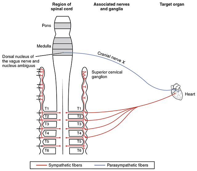
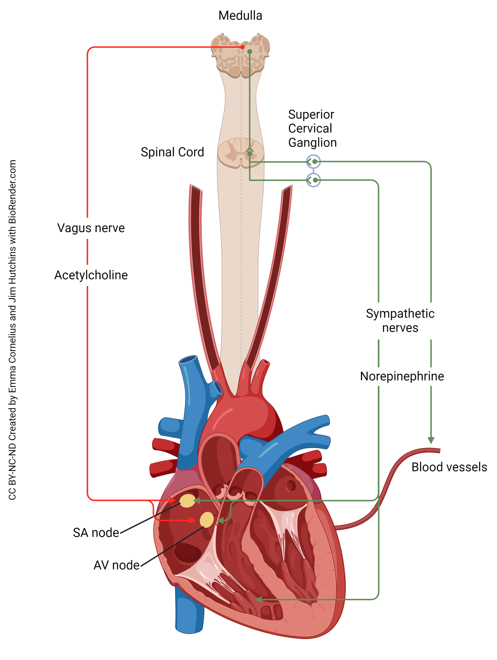
Recall that cranial nerve X, the vagus nerve, is named this because it wanders all over the thoracic and abdominal cavities. Therefore, cell bodies of the parasympathetic nervous system which innervate the heart are located in the dorsal motor nucleus of the vagus and the nucleus ambiguus. Their axons travel through the vagus nerve to reach the heart. Parasympathetic (“rest and digest”) action is to slow the heart, and it does this by releasing acetylcholine onto the pacemaker cells in the sinoatrial node of the heart.
For the sympathetic innervation of the heart, preganglionic cell bodies are found in the lateral horn of the spinal cord (green region of the diagram) at upper thoracic levels (there is some variability, but T1-T5 would be a good estimate). These send out axons which synapse in the T1-T5 sympathetic chain ganglia but also in some chain ganglia not directly linked to the spinal cord: the superior cervical ganglion, middle cervical ganglion, and inferior cervical ganglion. All these ganglia contain postganglionic cell bodies of neurons whose axons travel in the cardiac accelerator nerve to the pacemakers of the heart, where they release norepinephrine (noradrenaline) to speed up the heart. This is exactly what you want to do in a sympathetic (“fight or flight”) response.
The Effect of Norepinephrine on the Heart
[insert beta1 and beta2 transduction diagrams here]
Norepinephrine acts on the heart primarily through β-adrenergic receptors. Recall that these receptors are G protein-coupled receptors. This receptor type has a stimulatory Gα subunit (Gsα). When norepinephrine (or epinephrine, or noradrenaline, or adrenaline) bind to the receptor, a shape change causes the release of the Gsα which in turn bangs into, and stimulates, adenylyl cyclase (also called adenylate cyclase or AC). Adenylyl cyclase catalyzes the formation of second messenger cyclic AMP (cAMP). Cyclic AMP, in turn, increases the activity of the enzyme protein kinase A (PKA), which modifies a large number of intracellular proteins. The action of cAMP is terminated by an intracellular enyzme called a phosphodiesterase (PDE) which breaks the bond of phosphate to itself and turns cAMP into plain ol’ AMP.
[replace this diagram with a new one]
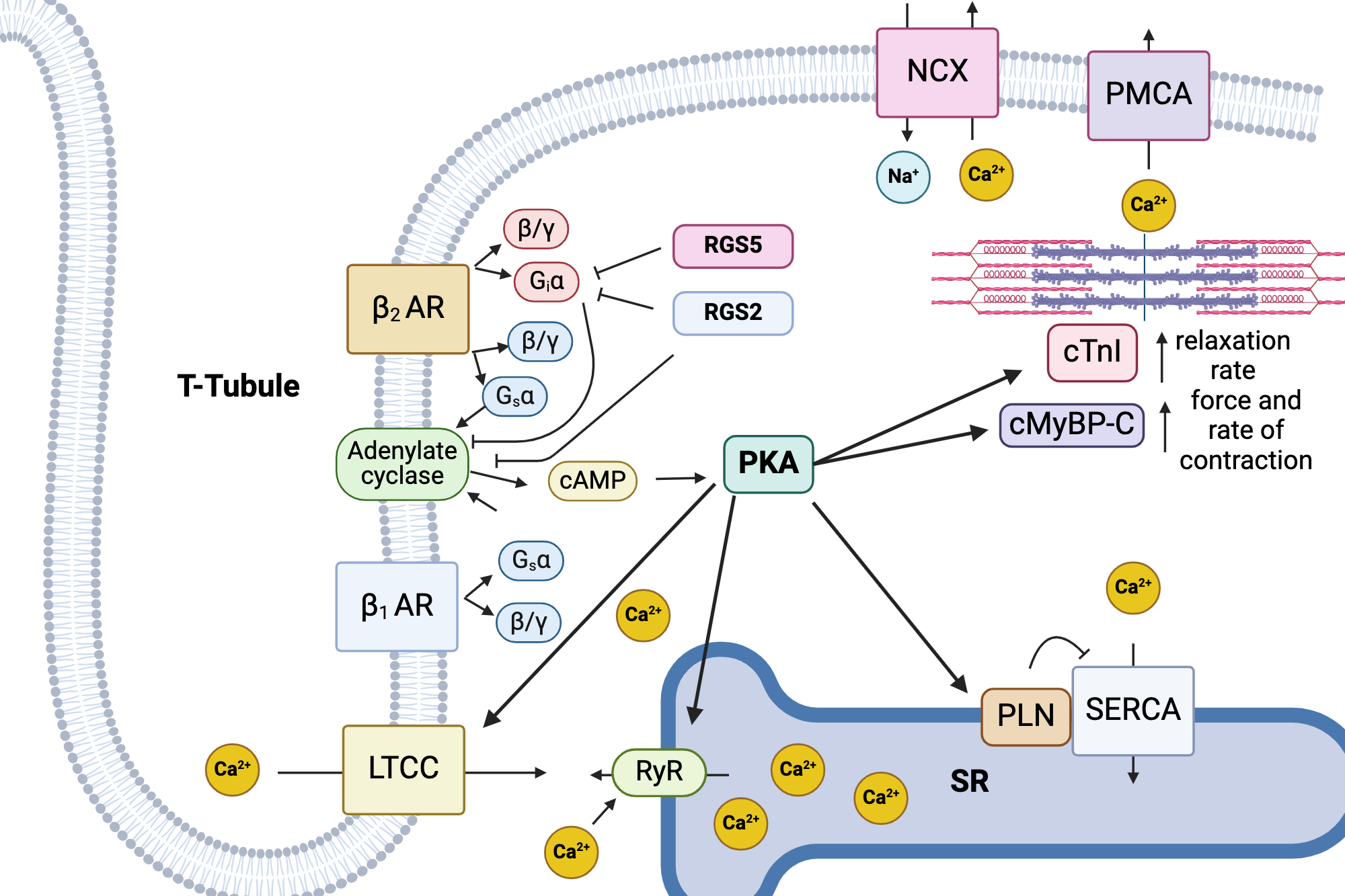
More recent evidence indicates that there is a much more complicated response inside the cardiac pacemaker and atrial cells; Ca2+ release from intracellular stores, stimulated by protein kinase A (PKA), plays a huge role in heart contractility. An additional release mechanism is an L-type Ca2+ channel (LTCC) . Both pathways stimulate a ryanodine receptor (RyR) on the sarcoplasmic reticulum which will then trigger Ca2+ release.
Media Attributions
- Autonomic Obj4Fig1 U12-026 heart autonomics v2.jpg © OpenStax is licensed under a CC BY (Attribution) license
- Autonomic Obj4Fig2 U12-025 Autonomic Innervation of the Heart v2 2172 px © Emma Cornelius and Jim Hutchins | BioRender is licensed under a CC BY-NC-ND (Attribution NonCommercial NoDerivatives) license
- Autonomic Obj4Fig2a biorender dahlen cardiomyocyte excitation-contraction coupling © Shelby Dahlen | BioRender is licensed under a CC BY-NC-ND (Attribution NonCommercial NoDerivatives) license
About the author
Dr. Jim Hutchins is an adjunct instructor at Colorado School of Mines with 45 years of teaching experience spanning K-12 through medical school. He earned his PhD in Neuroscience from Baylor College of Medicine, where his dissertation focused on acetylcholine as a neurotransmitter in the human retina, followed by postdoctoral research at Vanderbilt University on visual system development. His research contributions include highly cited work on mitochondrial superoxide dismutase and synaptic pruning in the retinogeniculate system. Dr. Hutchins has authored multiple open-access textbooks in neuroscience, medical terminology, and anatomy & physiology, creating freely accessible educational resources that have saved students over $5 million in textbook costs. He is committed to open educational resources and Creative Commons licensing, believing that knowledge should be freely available to all learners.
